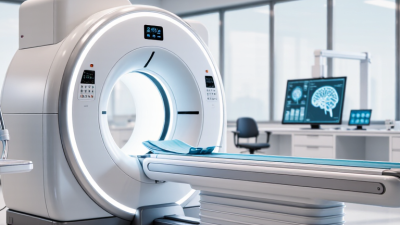
Choosing the right Neurology Machine can shape diagnosis, treatment planning, and daily clinical workflow. Hospitals need more than impressive specifications. They need dependable signals, clear images, simple operation, and responsive technical support.
Neuroscientist David Eagleman observes, “The brain is the most complex object in the known universe.” That complexity explains why neurological equipment must be evaluated carefully. A modern EEG system may capture subtle seizure activity beside a patient’s bedside. An EMG machine can help assess nerve conduction during a focused outpatient examination. MRI and CT systems offer different strengths, especially when speed, tissue detail, or emergency access matters.
This guide reviews seven leading neurology machines for hospitals and clinics in 2026. It considers diagnostic accuracy, patient comfort, software integration, maintenance demands, and practical return on investment. It also examines how each system fits different clinical environments, from large neurological centers to smaller community clinics.
Specifications can mislead.
A higher field strength does not automatically create better patient outcomes. More automated software may improve efficiency, but it can also hide important limitations. Staff training, calibration, room design, and reliable data storage remain essential.
The best choice is rarely the most expensive machine. It is the system that clinicians can use confidently, patients can tolerate safely, and administrators can maintain consistently. Some comparisons remain imperfect because performance depends on protocols and operator experience. That uncertainty deserves attention, not marketing language.
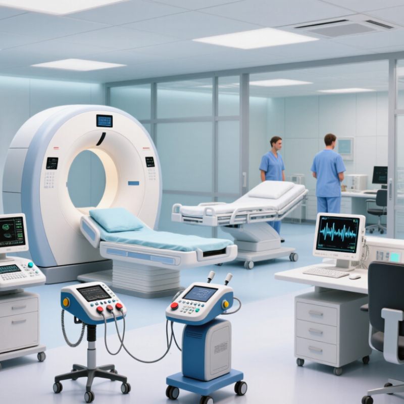
Neurology machines convert hidden nerve activity into measurable clinical evidence. Electroencephalography records electrical brain waves during seizures, sleep studies, or unexplained confusion. Electromyography and nerve conduction systems assess muscle response and peripheral nerve speed. Magnetic resonance imaging reveals tumors, strokes, inflammation, and spinal damage without ionizing radiation. Computed tomography offers rapid imaging when minutes matter. Evoked-potential systems measure sensory pathways, while intraoperative monitoring helps protect neural structures during complex procedures. Polysomnography examines brain, breathing, and muscle signals overnight.
The need is substantial. A 2024 analysis in The Lancet Neurology, using Global Burden of Disease data, estimated that 3.4 billion people experienced a neurological condition in 2021. These disorders caused about 443 million disability-adjusted life-years. The World Health Organization also identifies neurological care as a major health-system priority. Reliable machines support earlier triage, clearer diagnosis, treatment monitoring, and safer referrals. They do not replace clinical judgment. Poor electrode placement, motion, weak calibration, and rushed interpretation can still mislead a team. That uncomfortable limitation deserves attention.
Tips: Match each machine to local case volume, staffing, and emergency needs. Verify maintenance records, calibration schedules, data security, and clinician training. Ask for independent accuracy evidence, not only sales claims. A modern system can still underperform in a crowded room with insufficient technical support. Clinics should also plan backup workflows when imaging access or specialist review is delayed.
Choosing among the best neurology machines for hospitals and clinics in 2026 requires more than comparing features. Start with the clinical question. An EEG system needs stable signal capture, flexible electrode support, and clear artifact removal. An EMG unit should provide precise stimulation control and comfortable patient contact. For evoked-potential testing, timing accuracy matters greatly. Ask neurologists, technicians, and biomedical engineers to test each device during realistic examinations.
Look closely at daily workflow. Can staff create a patient record quickly? Are waveforms readable under dim clinic lighting? Does the system export reports in standard formats? Check electrode impedance, sampling performance, calibration steps, and battery behavior during a long session. A five-minute setup delay becomes significant in a busy outpatient department. Infection-control design also matters. Smooth surfaces and removable accessories simplify cleaning between patients.
Do not judge equipment by technical specifications alone. Review service response times, training quality, warranty terms, software updates, and replacement-part access. Verify electrical safety and applicable medical-device standards with qualified professionals. Protect patient data through role-based access and secure storage. A machine may perform well in a demonstration but struggle with tremor, movement, or electrical interference. That weakness deserves attention. No scorecard is perfect. I would document failed tests, staff concerns, and maintenance needs before approving a purchase. Price still matters, but unreliable recordings cost more through repeated examinations and delayed clinical decisions.
A comparison of three field strengths used in clinical MRI. The 7 T category represents specialized systems; availability and approved uses vary by region and facility.
Field strength alone does not determine which system is best for a patient or clinic. Evaluate it alongside clinical protocols, coils, image quality, artifacts, workflow, and service requirements.
Seven Leading Neurology Machines for Hospitals and Clinics in 2026
Neurology services in 2026 rely on several complementary machines, not one universal solution. Magnetic resonance imaging provides detailed views of the brain, spinal cord, and vascular structures without ionizing radiation. Computed tomography remains valuable during suspected stroke, trauma, or sudden bleeding because it works quickly. Electroencephalography records brain-wave activity and supports the assessment of seizures, altered awareness, and sleep-related disorders.
Electromyography with nerve conduction testing examines muscle response and peripheral nerve function. It can help investigate weakness, numbness, and suspected nerve compression. Polysomnography monitors breathing, oxygen levels, movement, and brain activity during sleep. Transcranial Doppler measures blood-flow velocity through cranial vessels and can support vascular monitoring. Positron emission tomography adds metabolic information, which may assist selected epilepsy, dementia, and tumor evaluations.
Practical selection requires more than impressive specifications. A busy hospital may prioritize scanning speed, emergency access, and integration with electronic records. A smaller clinic may need compact equipment, simple cleaning procedures, and dependable technical support. Staff training matters. So does calibration.
No machine is perfect. Motion can blur images, electrodes can lose contact, and test results may conflict with symptoms. Experienced clinicians should review findings alongside neurological examinations and patient history. Maintenance records, safety checks, and transparent reporting strengthen reliability. Some facilities may purchase advanced systems before building adequate training capacity. That is an avoidable weakness, but it still happens.
| Rank | Neurology Machine | Primary Clinical Purpose | What It Measures | Typical Clinical Configuration | Main Applications | Key Advantages | Important Considerations |
|---|---|---|---|---|---|---|---|
| 1 | Digital EEG System | Recording and analyzing electrical activity generated by the brain. | Scalp-recorded brain-wave activity, including background rhythms, epileptiform discharges, and seizure patterns. | 19 or more scalp electrodes Digital video Routine and prolonged recording | Epilepsy assessment, seizure classification, encephalopathy, altered consciousness, intensive-care monitoring, and pre-surgical evaluation. | Non-invasive, widely established, and suitable for routine, ambulatory, and video-EEG examinations. | Requires correct electrode placement, low electrical noise, artifact recognition, and interpretation by trained personnel. A normal routine EEG does not exclude epilepsy. |
| 2 | EMG and Nerve Conduction System | Assessing peripheral nerves, neuromuscular junctions, and skeletal muscles. | Nerve conduction velocity, latency, amplitude, sensory and motor responses, and needle-recorded muscle activity. | Motor and sensory studies Surface electrodes Needle EMG capability | Polyneuropathy, radiculopathy, entrapment neuropathy, plexopathy, motor neuron disease, and selected muscle disorders. | Provides complementary physiologic and anatomical information and can help localize lesions along the peripheral nervous system. | Needle examination is invasive and operator-dependent. Results are affected by temperature, patient age, technical settings, and the timing of disease. |
| 3 | Polysomnography System | Comprehensive diagnosis of sleep-related breathing and movement disorders. | Sleep stages, brain activity, eye movements, muscle tone, airflow, respiratory effort, oxygen saturation, heart rate, and body position. | Overnight recording Multi-channel monitoring Synchronized video | Obstructive sleep apnea, central sleep apnea, parasomnias, narcolepsy assessment, periodic limb movements, and complex sleep-related events. | Combines neurologic, respiratory, cardiac, and behavioral signals in one synchronized study. | Requires a controlled sleep environment and trained scoring staff. Sensor displacement, poor sleep quality, and first-night effects can influence results. |
| 4 | Intraoperative Neurophysiological Monitoring System | Monitoring nervous-system function during procedures that may place neural structures at risk. | Somatosensory evoked potentials, motor evoked potentials, spontaneous EMG, triggered EMG, EEG, and auditory or visual evoked responses when indicated. | Multi-modality recording Real-time alerts Operating-room integration | Spine surgery, intracranial procedures, vascular surgery, tumor resection, correction of spinal deformity, and selected ear or skull-base procedures. | Can provide continuous functional feedback while surgery is in progress and support preservation of motor, sensory, and cranial-nerve pathways. | Interpretation must account for anesthesia, blood pressure, temperature, muscle relaxants, electrical interference, and surgical factors. It requires a qualified monitoring team. |
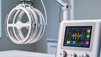
| 5 | Transcranial Magnetic Stimulation System | Non-invasive stimulation and functional assessment of cortical motor pathways. | Motor-evoked potentials, cortical excitability, motor threshold, and response to repetitive stimulation protocols. | Magnetic pulse generator Coil-based stimulation EMG integration | Motor pathway assessment, selected research protocols, and treatment of certain approved neuropsychiatric conditions under appropriate clinical guidance. | Does not require surgical access and can provide information about corticospinal tract function and cortical excitability. | Strict screening is required for seizure risk, implanted electronic devices, and metal near the head. Protocols, treatment indications, and safety limits must follow current clinical guidance. |
| 6 | Transcranial Doppler Ultrasound System | Evaluating blood-flow velocity and hemodynamics in the major intracranial arteries. | Blood-flow velocity, direction, pulsatility, embolic signals, and changes during physiologic or therapeutic maneuvers. | Pulsed-wave Doppler Color-flow option Monitoring probes | Vasospasm surveillance after subarachnoid hemorrhage, emboli detection, stroke assessment, sickle-cell screening, and cerebrovascular reactivity studies. | Bedside, repeatable, radiation-free, and capable of continuous monitoring in selected patients. | Results depend on operator skill and acoustic-window quality. Flow velocity is not the same as blood-flow volume and should be interpreted with clinical and imaging findings. |
| 7 | Evoked Potential System | Testing the functional integrity of sensory pathways from peripheral receptors to the cerebral cortex. | Latency and amplitude of responses to visual, auditory, or somatosensory stimulation. | VEP capability AEP/BAEP capability SSEP capability | Multiple sclerosis evaluation, optic-pathway assessment, hearing-pathway testing, spinal-cord assessment, coma evaluation, and intraoperative monitoring. | Can identify conduction delay or pathway dysfunction that may not be evident from the clinical examination alone. | Responses are sensitive to attention, temperature, age, visual acuity, hearing status, sedation, electrode placement, and technical artifact. |
MRI remains a leading neurological imaging system in 2026. It provides detailed views of brain tissue, vessels, tumors, and spinal structures without ionizing radiation. Diffusion and functional sequences improve stroke and surgical planning. However, scanning is expensive, slow, and difficult for patients with severe anxiety or some implants.
CT delivers rapid images during suspected bleeding, trauma, or acute stroke. Its strengths include wide availability and excellent bone detail. Radiation exposure remains a concern.
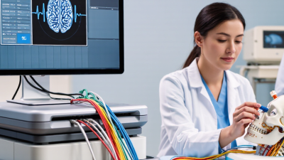
EEG records electrical brain activity through scalp electrodes and supports epilepsy, coma, and sleep evaluations. It is portable and relatively affordable. Yet, movement, muscle activity, and poor electrode contact can distort results.
Electromyography and nerve conduction systems assess peripheral nerves, weakness, and suspected neuropathy. They offer useful functional data, but testing may feel uncomfortable and depends heavily on operator technique.
PET shows metabolic changes linked with dementia, epilepsy, and selected tumors. It requires radioactive tracers, specialized handling, and careful interpretation.
Transcranial Doppler measures blood flow through cranial vessels and can support vasospasm monitoring. Its limitation is an acoustic window that some patients lack.
Intraoperative neurophysiological monitoring tracks neural pathways during complex surgery. It can warn teams about functional changes, but anesthesia and technical errors may affect signals.
No machine replaces clinical judgment. Availability, maintenance, staff training, and local patient needs should shape the final choice.
Choosing among the 7 best neurology machines starts with the patients and tests your facility actually serves. An EEG system suits seizure assessment and brain-wave monitoring; EMG and nerve-conduction equipment supports evaluation of muscle and peripheral nerve function. Evoked-potential systems may be useful when clinicians need to measure responses to specific sensory stimuli. One machine rarely covers every need.
Check more than the feature list. Confirm that recording channels, sampling rates, electrodes, and software fit your protocols. Ask staff to test the controls, patient leads, and report workflow before purchase. A cluttered interface can slow a busy clinic. So can awkward cable storage. Consider room size, cleaning procedures, training, warranty coverage, and access to qualified service technicians. These details affect daily use, not just the initial setup.
Compare the full cost of ownership, including accessories, maintenance, and staff training. Request a demonstration using realistic cases, then have neurologists and technicians assess image or signal quality and ease of use. Verify that the device can export data in formats your records system accepts. Specifications can look excellent on paper. They do not guarantee a smooth clinic day. Be candid about what your team has not tested yet, and leave room to revise the shortlist.